 |
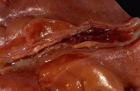
| Figure 1.0 Deposition of yellow fatty streaks in the artery. |
Cholesterol is a lipid which is an essential structural component of mammalian cell membranes and is required to establish proper membrane permeability and fluidity. In addition, cholesterol is an important component for the manufacture of bile acids, steroid hormones, and vitamin D.
 |
| Figure 1.1 HDLs have a lower content of cholesterol |
Cholesterol is insoluble in water and is transported in the blood plasma in the form of tiny balls of lipid and protein called lipoproteins. There are 2 groups of lipoproteins , low density lipoproteins (LDL) and high density lipoproteins (HDL).
LDLs transport cholesterol from the liver to the tissues, including the artery walls. They tend to deposit their cholesterol at any damaged sites. However, HDLs remove cholesterol from tissues and transport it to the liver to be excreted. Hence, HDLs help to protect arteries against atherosclerosis.
 |
| Figure 1.2 White blood cells stick to the endothelium. |
When endothelium cells sense an injury, they produce signals that prompt smooth muscle cells in the arterial wall to change.
Thus, the smooth muscle cells moving toward the site of vascular injury, where they reposition themselves just beneath the endothelial cell layer.
In reaction to injury, endothelial cells also produce substances that signal circulating blood cells to stick to the endothelium instead of flowing through the vessel.Lipid or fat cell-like substances in the blood, such as LDLs and triglyceride will then accumalate in this area.
The lipids that accumulate in the broken endothelium become oxidized. The endothelium cells were sent signals, which then alert the smooth muscle cells to begin a ''repair'' process that eventually results in an atherosclerosis lesion.
 |
| Figure 1.3 Plaque forms in the artery. |
These white blood cells and smooth muscle cells, which are called ''foam cells'', induce chronic inflammatory attack by various immune components. Smooth muscle cells try to curtail the injury to the endothelium by producing collagen which forms a cap over the injury site.
Then calcium accumulates and forms a material resembling bone. These complex array of foam cells, calcification and lipid accumulation is called a plaque.
As a plaque increases in size, it protrudes into the lumen of the artery and begins to block it. This commonly occurs in the aorta and coronary arteries which supply the oxygen to the muscle of the heart.
 |
| Figure 1.4 Artery begins to block as the plaque increases in size. |
The artery wall is made weaker by atheromatous plaques and may stretch as a result. Local stretching is called aneurysm.
If it ruptures, the process is known as haemorrhage. This is more likely if atherosclerosis has occurred.
If it ruptures, the process is known as haemorrhage. This is more likely if atherosclerosis has occurred.
Once an artery is blocked, the tissue it supplies will lack of oxygen and will be severely damaged or die. If thrombosis occurs in a coronary artery (coronary thrombosis), the heart is damaged and a ‘heart attack’ may occur. The medical term used for a heart attack is myocardial infarction. If thrombosis occurs in the brain (cerebral thrombosis), a stroke may occur.
 |

No comments:
Post a Comment